Imaging Zebrafish Larvae to Evaluate Future Drugs
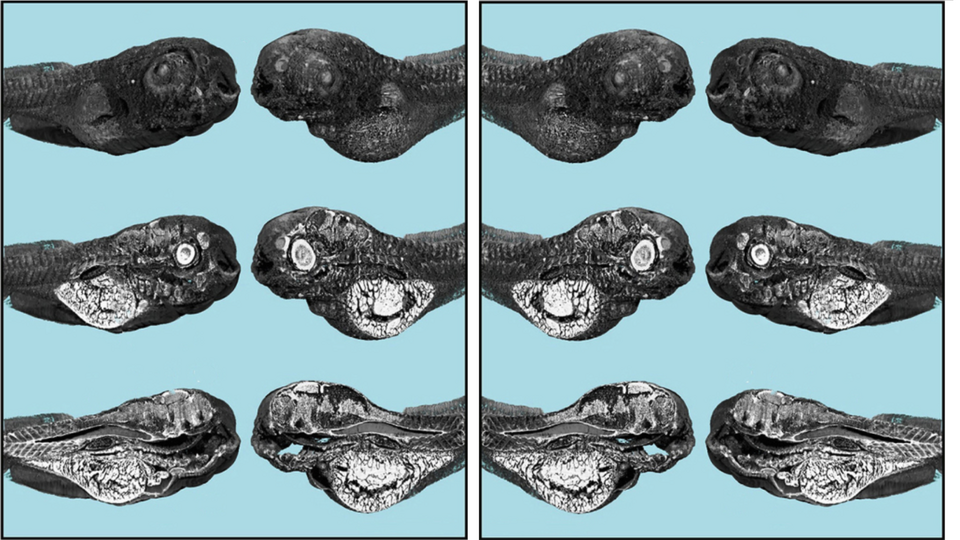
Future drugs must be tested to see if they harm the kidney. Cell-based assays are often used for this purpose, although it is difficult to extrapolate the results to vertebrates. Researchers of the Biomaterials Science Center and the MiNa Core Facility together with pharmaceutical technologists and French beamline scientists, therefore, have evaluated whether zebrafish larvae could serve as a vertebrate screening model to detect changes of renal organs induced by the antibiotic gentamicin.
Zebrafish larvae are well suited to study renal morphological aberrations and are a predictive model for acute human kidney injury since the functional unit of their kidney closely resembles the human anatomy. Therefore, they can be used as an alternative to animal experiments in higher vertebrates according to the 3R principles of animal welfare.
To visualize gentamicin-induced renal toxicity, Bert Müller and his team have employed synchrotron radiation-based micro computed tomography (SRµCT) at the ANATOMIX beamline of synchrotron SOLEIL. This method provides 3D representations of renal structures with true micrometer resolution, without the need to label the organs studied.
The scientists conclude that zebrafish larvae may be suitable as an intermediate model to assist in the extrapolation from cell-culture-based test systems to mammals.
The approach can therefore be used to identify potential nephrotoxins in drug discovery or environmental sciences.
More:
- The related paper: Bolten, J.S., Tanner, C., Rodgers, G., Schulz, G., Levano, S., Weitkamp, T., Waldner, S., Puligilla, R.D., Bodmer, D., Müller, B., Huwyler, J. "Zebrafish (Danio rerio) larvae as a predictive model to study gentamicin-induced structural alterations of the kidney" PLoS One., 18(4): art.n° : e0284562. (2023).
- An article on the website of the Soleil Synchrotron
- Biomaterials Science Center
- Core Facility MiNa