Editor’s pick: Optics and the Brain
Three-dimensional brain imaging is generally performed with magnetic resonance tomography. X-ray tomography, however, provides substantially improved spatial resolution. Replacing the established absorption contrast by the phase-contrast modality, researchers succeeded in the visualization of the cells in the human brain. The next chapter is human cerebellum tomography with near-field speckles, published by an international team with Christos Bikis and Bert Müller from the Biomaterials Science Center at the University of Basel.
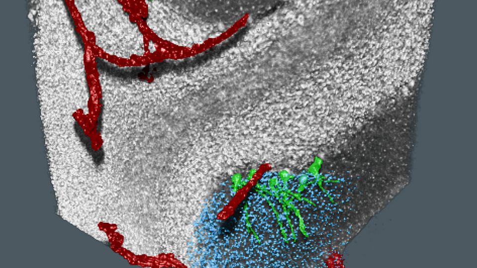
The working principle of speckle-based imaging relies on the addition of a randomly structured mask, such as sandpaper, into the beam to generate a speckle pattern. This pattern changes as X rays travel through the sample, encoding distinct physical properties of the tissue under investigation. The interdisciplinary research team has used this approach for the non-destructive, multi-modal inspection of paraffin-embedded human brain tissue. The three-dimensional images show enough contrast and spatial resolution to not only make visible the Purkinje cells but also to locate the stellate and granule cells within the human cerebellum. In addition, the dark-field modality provides insights on the unresolved microstructure within the cerebellar tissue.
More: